Pregnancy in Dubai

Advice & information on common pregnancy problems by a Fetal & Maternal Medicine Consultant


1 Comment

Nuchal translucency and screening for Down’s syndrome in Dubai.

What is nuchal translucency ( NT ); The term nuchal translucency is used to describe the sonographic (ultrasound scan) appearances of a specific region of the baby’s neck during the first trimester of pregnancy. It is routinely measured between weeks 11 -13+6 days and used as a screening test for chromosomal abnormalities like Down’s (trisomy 21).
What do you need to know about the nuchal translucency;
The quality of Nuchal translucency measurement (and its prediction accuracy) depends on who is doing it and what equipment is used. If you decide to have the test done ensure it is performed by properly trained and certified operators that use good quality equipment.  One of the well respected certification organisations is The Fetal Medicine Foundation. You will find in this link lists of certified professionals for your country.
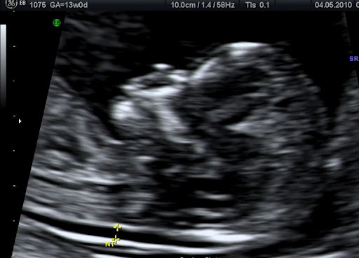
Normal Nuchal Translucency

NT is only a part of the first trimester scan. Although commonly referred as NT scan at the first trimester scan the doctor / sonographer should report on how some basic features of our baby have developed as well. For example the fetal abdomen the baby’s skull and brain, the fetal stomach and bladder the arms and legs should be clearly visible in most cases.

Nuchal translucency is a powerful predictor for problems like Down’s syndrome. It is routinely used in combination with maternal age and biochemical markers (blood test) to give a risk estimate for major chromosomal anomalies (T21, T18, T13).
The size of the nuchal translucency depends on the gestational age (how old the baby is). The upper limit of normal is smaller at 11 weeks and larger at   13 weeks.
Ultrasound scanning measurement of the NT is only a screening and not a diagnostic test. This means that it will provide you with a risk estimate rather than a “Yes – No” answer.
It is best practice to discuss with your obstetrician and understand what is the NT scan beforehand. Make sure you have some quality time with your doctor to understand the purpose of the test, the results you expected to have as well as the limitations it has.


Dr George Michailidis, Obstetrician and Gynecologist 
Fetal and Maternal Medicine Consultant 


Leave a comment

Having a dating ultrasound scan in Dubai

The common way to calculate how far your pregnancy has progressed is to use Naegele’s rule. This simple equation uses the first day of your last period it adds 9 months and 7 days. This is on what the small plastic wheels that you usually see in antenatal clinics are based.
As you can understand although in general this works well there a number of weak points.
1.The mother may not be certain of the first day of the last period.
2.The rule assumes that each mother has regular periods and produces an ovum (egg) on day 14 from the first day off the period.
It is easy to appreciate that this is not always the case. Humans are not like Swiss watches and there are normal variations from month to month.
Accurate dating of the pregnancy is important as it constitutes the starting point for planning pregnancy care.
The most accurate way to date your pregnancy (with the exception of IVF pregnancies) is the dating ultrasound scan. This is usually performed between weeks 8-10.
The purpose of the dating scan is
  1. To confirm a viable (alive) intrauterine (within the womb) pregnancy.  This is achieved by demonstrating a beating fetal (baby) heart.
  2. To date the pregnancy. This is achieved by measuring how long the fetus is from the top of its head to its bottom  (Crown Rump Length – CRL). Between 8-10 weeks it is the best time to calculate how far you are in your pregnancy (accuracy about 4 days).
  3. To diagnose multiple pregnancies. Diagnosing twins or higher order multiple pregnancies early helps appropriate pregnancy planning.
Most of the times the scan is done transabdominally. This means the probe that produces and receives ultrasound waves touches your lower abdomen ( tummy) and allow us to get views  of the uterus (womb). Special gel is used to improve contact between the probe and your tummy.
In some occasions a transvaginal (internal) scan is needed.

This means that a special probe is inserted in the vagina. In this way it gets much closer to the womb and produces a much clearer image. This is more commonly needed in very early pregnancy, for those women who are overweight or have a backwards tilted womb.

A good doctor should explain to you the type of scan that needs to be done and a chaperone should will be in the room during internal scans.
Having a scan should not be painful. If in any case you feel uncomfortable let the doctor know as he/she is usually concentrating at the ultrasound screen.
What information will I get ?
It is best obstetric practice to give a report which summarizes the scan findings at the end of the examination ie; position of the pregnancy, number of fetuses (babies), fetal heart activity, CRL and the calculated gestation and expected date of delivery.
At this stage it unlikely due to the very small size of the baby that even severe abnormalities will be picked up.
Make sure you keep a copy of the report as the expected date of delivery should not be changed again during the pregnancy. Providing the initial scan has been performed by a well trained doctor, good obstetricians know well that follow up scans should confirm how your baby is growing against the already set date and not change the dates. So when you see a new doctor bring that documentation with you as it will help with your pregnancy management.
Can the dating scan pick up any problems?
 At this stage of the pregnancy possible problems that can be picked up are
  1. A missed miscarriage. This is when the pregnancy has stopped developing and there is no visible fetal heart beat. A miscarriage in early pregnancy unfortunately is not uncommon. Most of the times no specific reason can be identified. Your gynecologist will explain the findings to you and arrange with you the appropriate management.
2.A pregnancy cannot be clearly seen within the womb. Possible reasons for this are; a very early pregnancy, a miscarriage, or in some occasions an ectopic pregnancy. Your Gynecologist  will explain the findings to you and initiate the appropriate investigations needed to clarify the diagnosis. In those cases that an ectopic pregnancy is diagnosed or strongly suspected an operation might be needed to deal with it.

Dr George Michailidis, Obstetrician and Gynecologist 
Fetal and Maternal Medicine Consultant 


3 Comments

First Trimester Ultrasound Scan (11-13 weeks)

What is the purpose of the first trimester scan?

The purpose of this scan is;

1.     To confirm a viable (alive) intrauterine (within the womb) pregnancy.  This is achieved by demonstrating a beating fetal (baby) heart.
2.     To date the pregnancy. This is achieved by measuring how long the fetus is from the top of its head to its bottom (Crown Rump Length – CRL accuracy about 7 days).
3.     To diagnose multiple pregnancies. Diagnosing twins or higher order multiple pregnancies early helps appropriate pregnancy planning.
4.     To provide a basic anatomical survey of the developing baby. A number of common major structural anomalies can be seen at this stage.
5.     If you wish in addition to the anatomical survey a number of specialist measurements can be done to help calculate the risk of your baby being affected by chromosomal anomalies like Down’s syndrome.

How is the scan done?

Most of the times the scan is done transabdominally. This means the probe that produces and receives ultrasound waves touches your lower abdomen (tummy) and allow us to get views  of the uterus (womb). Special gel is used to improve contact between the probe and your tummy.
In some occasions a transvaginal (internal) scan is needed. This means that a special probe is inserted in your vagina. In this way it gets much closer to the womb and produces a much clearer image. This is more commonly needed in very early pregnancy, for those women who are overweight or have a backwards tilted womb.
The doctor will explain to you the type of scan that needs to be done and a chaperone will be in the room during internal scans.
Having an ultrasound scan should not be painful. If in any case you feel uncomfortable let the doctor know as he/she is usually concentrating at the ultrasound screen.
What about having a full bladder? A full bladder helps with transabdominal scans as it pushes your womb upwards and at the same time pushes the bowel out of the way creating good window to look at your baby. However you should not feel uncomfortable and be able to enjoy your scan. 

What information will I get?


At the end of the scan you will be given a printed report which will summarize the scan findings ie; position of the pregnancy, number of fetuses (babies), fetal heart activity, CRL and the calculated gestation and expected date of delivery. Basic anatomical structures that can be seen at this stage will also be described.
At this stage due to the very small size of the baby only specific severe abnormalities will be picked up. Typical problems picked up at this stage are acrania (when the fetal skull has not develop), gastroschisis and omphalocele (anomalies that affect how the baby’s tummy has formed). Futhermore as the baby continues to develop further, some problems are evident later in the pregnancy.
Problems with the baby’s heart are not commonly seen at this stage. The main scan aiming to reassure that baby is structurally normal is the 20 week scan.
The baby’s gender is not easy to be determined at this stage. Male and female external genital organs look very similar at this gestation. However an educated guess (about 80-90% accurate) can be attempted if the views are good.

What are the risks for my pregnancy?

Ultrasound scanning has been routinely used in obstetrics for more than 20 years. Routine scanning does not pose any risk to your baby or yourself. 
What if there is a problem?
Most scans are happy experiences but sometimes problems are either seen or suspected. At this stage of the pregnancy problems that can be picked up are
1.     A missed miscarriage. This is when the pregnancy has stopped developing and there is no visible beating fetal heart. A miscarriage in early pregnancy unfortunately is not uncommon. Most of the times no specific reason can be identified. The doctor will explain the findings to you and arrange with you the appropriate management.
2.   Suspicion or evidence of a structural anomaly (problems relating to how the baby has developed). The doctor will explain the findings to you and depending on the problem explain what are the implications for you pregnancy and the baby. I some occasions further tests may be needed to clarify the diagnosis.
What if I have more questions ?

We hope you will find this information useful. If you have any questions you can contact either Dr Michailidis or our specialist midwife

Dr George Michailidis, Obstetrician and Gynecologist 
Fetal and Maternal Medicine Consultant