Pregnancy in Dubai

Advice & information on common pregnancy problems by a Fetal & Maternal Medicine Consultant


Leave a comment

Preterm birth and cervical length screening in Dubai

Preterm premature birth is the leading cause of neonatal death and long term morbidity. It has a huge impact in the affected families with both physical and psychological implications. From the provider health-care system point of view employing effective screening and treatment methods translates in major good outcome benefits including great health care savings.


The American College of Obestrics and Gynecology and the Society of Maternal Fetal Medicine have recently published their position on the issue of screening and prevention of preterm birth. 

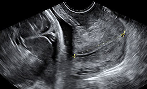
Both Societies  acknowledged the importance of cervical length as a predictor of preterm labor and the efficacy of vaginal progesterone in improving pregnancy outcomes 


In summary the some of the SMFM recommendations are the following
1 In women found to have a prematurely short cervix are at high risk for preterm labour and should be treated

2 Use of progesterone is associated with lower risk for preterm birth in some pregnant women, as well as with better neonatal outcomes


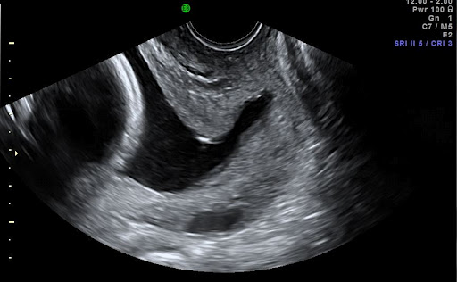
3 Because of the high prevalence and public health burden of preterm birth, experts recommend universal cervical length measurement, which meets World Health Organisation criteria for an effective screening test.

Preterm labor in Dubai there are additional issues that the family physician need to consider. The availability and of neonatal unit beds, the high costs of care for the baby, the rather fragmented after care. See our blog What you need to know about preterm labor in Dubai.

The best approach is prevention. A methodical approach including your detailed history and Cervical measurement done by qualified and experienced clinicians is likely to give the best results. Discuss with your obstetrician the best way to tackle preterm labour.  Feel free to contact us for further information.


Dr George Michailidis, Obstetrician and Gynecologist 
Fetal and Maternal Medicine Consultant 


Addtional resourses 


Berghella, V., The Society for Maternal-Fetal Medicine: 
Publications Committee, Progesterone and Preterm Birth Prevention: Translating Clinical 
Trials Data into Clinical Practice, American Journal of Obstetrics and 
Gynecology (2012), doi: 10.1016/j.ajog.2012.03.010.


1 Comment

Nuchal translucency and screening for Down’s syndrome in Dubai.

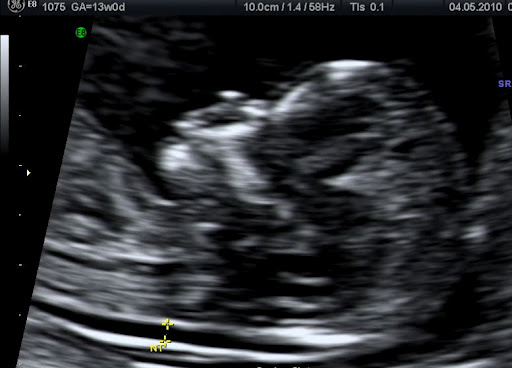
What is nuchal translucency ( NT ); The term nuchal translucency is used to describe the sonographic (ultrasound scan) appearances of a specific region of the baby’s neck during the first trimester of pregnancy. It is routinely measured between weeks 11 -13+6 days and used as a screening test for chromosomal abnormalities like Down’s (trisomy 21).
What do you need to know about the nuchal translucency;
The quality of Nuchal translucency measurement (and its prediction accuracy) depends on who is doing it and what equipment is used. If you decide to have the test done ensure it is performed by properly trained and certified operators that use good quality equipment.  One of the well respected certification organisations is The Fetal Medicine Foundation. You will find in this link lists of certified professionals for your country.

Normal Nuchal Translucency

NT is only a part of the first trimester scan. Although commonly referred as NT scan at the first trimester scan the doctor / sonographer should report on how some basic features of our baby have developed as well. For example the fetal abdomen the baby’s skull and brain, the fetal stomach and bladder the arms and legs should be clearly visible in most cases.

Nuchal translucency is a powerful predictor for problems like Down’s syndrome. It is routinely used in combination with maternal age and biochemical markers (blood test) to give a risk estimate for major chromosomal anomalies (T21, T18, T13).
The size of the nuchal translucency depends on the gestational age (how old the baby is). The upper limit of normal is smaller at 11 weeks and larger at   13 weeks.
Ultrasound scanning measurement of the NT is only a screening and not a diagnostic test. This means that it will provide you with a risk estimate rather than a “Yes – No” answer.
It is best practice to discuss with your obstetrician and understand what is the NT scan beforehand. Make sure you have some quality time with your doctor to understand the purpose of the test, the results you expected to have as well as the limitations it has.


Dr George Michailidis, Obstetrician and Gynecologist 
Fetal and Maternal Medicine Consultant 


Leave a comment

Having your 20 weeks ultrasound scan (morphology or anomaly scan) in Dubai; what you need to know.

Having your 20 weeks ultrasound scan (morphology or anomaly scan) in Dubai; what you need to know.

Ultrasound scanning has revolutionised obstetric practise. It provides a reliable and safe method of examining the developing baby.  However what it is not widely known in Dubai and in most countries is that formal ultrasound training only very recently became part of training in our speciality even in well respected western countries. As a result although most good obstetricians can perform a basic scan only a handful have the skills and experience to perform a full morphology scan. It is a huge misconception that a good doctor can do everything. It is common practise  for the best obstetricians to refer their clients to centres (like our Fetal Medicine Unit) which routinely perform ultrasound examinations.

What is the purpose of the 20 weeks ( morphology ) ultrasound scan?
The purpose of this scan is;
  1. To confirm a viable (alive) intrauterine (within the womb) pregnancy.  This is achieved by demonstrating a beating fetal (baby) heart.
  2. To provide a detailed anatomical survey of the developing baby. A number of major structural anomalies can be seen at this stage.
  3. To describe the position of the placenta (afterbirth).
If it happens to be the first scan in your pregnancy
  1. To date the pregnancy if this happens to be your first scan in your pregnancy. Although this is not as good as dating by an early pregnancy scan it still has an accuracy of about 7 days.
  2. To diagnose multiple pregnancies if this is our first scan. Diagnosing twins or higher order multiple pregnancies early helps appropriate pregnancy planning.

How is the scan done?

Most of the times the scan is done transabdominally. This means the probe that produces and receives ultrasound waves touches your lower abdomen (tummy) and allow us to get views of the uterus (womb). Special gel is used to improve contact between the probe and your tummy.
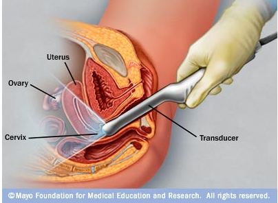
Very rarely a transvaginal (internal) scan is needed. This means that a special probe is inserted in your vagina. By approaching the baby from a different angle some structures may be seen better this way.
If however you opt to have your cervix(neck of the womb) measured as a screening test for preterm labour this is best done by an internal scan.
 The doctor will explain to you the type of scan that needs to be done and a chaperone will be in the room during internal scans.
Having an ultrasound scan should not be painful. If in any case you feel uncomfortable let the doctor know as he/she is usually concentrating at the ultrasound screen.
What about having a full bladder? A full bladder helps with transabdominal scans as it pushes your womb upwards and at the same time pushes the bowel out of the way creating good window to look at your baby. However you should not feel uncomfortable and be able to enjoy your scan.
Should I bring my friends and family with me? Although the scan is usually a very pleasant experience still the doctor need to concentrate as it is an important medical examination. In most cases it is best if your partner or a friend accompanies you at the scan room rather than a large number of people.   

What information will I get?

At the end of the scan YOU SHOULD BE GIVEN A COMPUTERIZED REPORT which will describe the structures seen and if there were any particular difficulties during the scan. THIS IS A VERY IMPORTANT PART OF YOUR ANTENATAL RECORD AND IT IS VERY USEFUL TO HAVE A COPY.
At this stage a large number of structural problems that a baby may have can be picked up. Ultrasound can usually provide information as to how various organs are put together and not how well they function.
Please note that scans are not foolproof. They do not show all the anomalies and in some occasions anomalies can be missed. A normal scan is not a guarantee of the delivery of a perfect baby.
Sometimes it is difficult to get good views of the baby. Common reasons for this is the baby’s position, an overweight mother or scar tissue from previous operations. In such cases the scan might need to be repeated another day.
Usually we will be able to comment on the baby’s gender at this stage. Please make sure that you let the doctor know if this is something that you want or do not want to know before the scan starts. Although doctors and sonographers are pretty good in finding the baby’s sex at this stage you should be aware that
  1. It is not 100% accurate
  2. It is not one of the primary aims of the scan (this is to confirm that major structures are normal) and it might not be possible to be seen during the allocated time. 

What are the risks for my pregnancy?

Ultrasound scanning has been routinely used in obstetrics for more than 20 years. Routine scanning does not pose any risk to your baby or yourself. 

What if there is a problem?

Most ultrasound scans are happy experiences but sometimes problems are either seen or suspected. At this stage of the pregnancy possible problems that can be picked up are
  1. Suspicion or evidence of a structural anomaly (problems relating to how the baby has developed). The doctor will explain the findings to you and depending on the problem explain what are the implications for you pregnancy and the baby. If you are having your scan by a sonographer they should be able to inform you of their concerns and arrange an urgent consultation with your obstetrician. In most cases your doctor after explaining the initial findings will arrange for you to see a Fetal Medicine Specialist. In some occasions further tests may be needed to clarify the diagnosis.
  2. A low lying placenta. Although this is not a problem at this stage of the pregnancy further scans will be needed later in pregnancy to confirm that the placenta has moved out of the way. Ask the doctor to provide you with the relevant information sheet.

  What if I still have questions about my scan?

Do not feel afraid to ask questions about aspects of the scan or the report that you do not understand. Your doctor should be able to provide you with clear information or arrange further consultation with a specialist.  

Dr George Michailidis, MRCOG
Fetal & Maternal Medicine Specialist
Consultant Obstetrician & Gynaecologist


4 Comments

What is a screening test? what is the difference from a diagnostic test?

(The following is an extract from our Fetal Medicine information sheet for Downs Syndrome.) 

Screening tests give as a result a risk estimate, in other words how likely it is for a person to have a certain condition. This is usually expressed as a numerical value for example 1:100. Many times for simplicity if the risk is higher than a certain value the test is reported as “high risk” or “low risk” if it is lower than the threshold selected. 

Screening tests do not confirm or exclude a condition. For example a screening test does not say that a baby “has” or “has not” Down’s syndrome.
They do however, provide valuable information to help the parents decide if they want to proceed with diagnostic tests.

Diagnostic tests are those that confirm or exclude a condition.
If the screening tests do not tell us “yes or no” why don’t we just have the diagnostic tests ?
Today the available diagnostic tests for Down’s syndrome are
1.Chorionic villus sampling CVS
2. Amniocentesis
Both tests carry a small risk of causing a miscarriage (0.5-1%).
For this reason many parents would opt to have first a screening test and use the results to help them decide if they would proceed to a diagnostic test.
What are the recommended screening tests?
In the past the main screening test for Down’s syndrome was the mother’s age. Unfortunately this was proven to be neither very sensitive (picked up about 60% of babies with T21) nor very specific (too many women had to have unnecessary invasive procedures).
 At the American Hospital Dubai we recommend;
1.First trimester combined test. This test combines the results of first trimester ultrasound scanning (Nuchal translucency, fetal heart rate) with biochemical markers (bHCG, PAPP A). The test is done at 11-13 weeks at the Fetal Medicine Unit of the American Hospital Dubai. The test detection rate is approximately 90%
2.Second trimester biochemistry. For those mothers who have booked late (after 13weeks and 6 days) we recommend the quadruple biochemistry test. This is a blood test for the mother. The detection rate is approximately 75%.
What is considered to be a high risk result?
The result of the test is considered to be “positive” if the risk estimate is higher than  1/300. For a example 1/240 is considered a “positive” or “high risk” result while 1/310 is considered a “negative” or “low risk” result.
It is always challenging for the parents to appreciate the magnitude of risk. 
Please take time to discuss the results with your Doctor as finally a good result is a result the parents are content with.

Dr George Michailidis, Obstetrician and Gynecologist 
Fetal and Maternal Medicine Consultant 


1 Comment

What are the questions a couple / mother in Dubai should think before embarking in testing for Down’s syndrome?

In pregnancy mothers undergo a large number of tests, many times without actually fully appreciating what each test is about. In particular screening and diagnostic tests for Downs syndrome T21 can end up causing a lot of anxiety. 
It would worth thinking and discussing with your partner and your doctor the following questions;

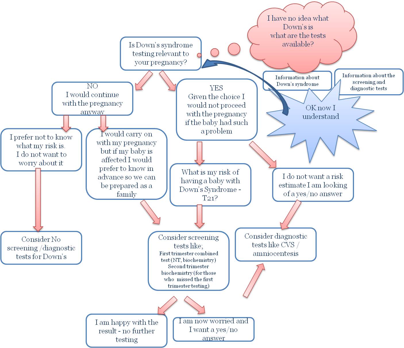
  1. Is this a relevant test?  In other words would it change the way you view this pregnancy?
  Usually parents do come in 3 big groups;
      Those for whom whether the baby has or not Downs make no difference what so ever. These parents would not consider termination and they would look after the baby the same way as any other baby would come their way.

       Those parents who believe that given the choice they would not bring to life a baby  with such a problem. 

       Those that say “Doctor I have no idea what I would do. Lets discuss what are the chances of this happening to help us make up our mind how will go forward”


For the first group testing for Downs may not be appropriate. It may result in extreme anxiety if the results are not reassuring without any real benefit for the overall pregnancy management.
I should point out here that although the majority of parents in this group would prefer not to know and worry, a small number would opt to undergo testing as they would like to be prepared (psychologically, making appropriate family arrangement etc..)


In the second group usually we see a number of parents that are looking for absolute certainty. They do not want a risk estimate but a “yes or no” answer to help them early make arrangement for the pregnancy management. For these parents  diagnostic tests like a CVS or an amniocentesis should be considered.


The third group contains the majority of parents and they should try and think the following;

  1. Is there a risk level that will make you feel content and secure?
    Unfortunately the results of screening tests give a risk estimate. The cut off points are usually set anywhere between a risk of 1/250 to 1/300. So a result 1/299 – abnormal – high risk result is statistically not different from the 1/301 – normal- low risk result. Dealing with the results of the test is challenging for everybody and you should spend some time with your doctor  discussing these issues.
  2. Avoid the question “doctor what would you do in my case?“. Each family has a unique setting. Even within the same family the circumstances change over a period of time. The role of you doctor in most of the cases should not be to tell you what to do but to give the information you need to help you make up your mind what is right for your family.   

The following flow chart is summarising the decision making process
Dr G Michailidis 
Make sure you have some quality time with your doctor and discuss the issues above. 

Remember for most of these questions there are no universally “right” answers. The correct plan is the one that is appropriately tailored to you and your family.

Dr George Michailidis, Obstetrician and Gynecologist 
Fetal and Maternal Medicine Consultant