Pregnancy in Dubai

Advice & information on common pregnancy problems by a Fetal & Maternal Medicine Consultant


Leave a comment

Flu vaccination during pregnancy in Dubai

The American College of Obstetrics and Gynecology has issued a new guideline advising women to get the flu vaccine. Traditionally, routine medical advice has been for women to shy away from all non essential medications during pregnancy. This has never changed what need however to change is the perception of the public about the potential consequences of sever flu during pregnancy and the safety   of the vaccines.
The 2009 H1N1, swine flu virus pandemic has brought to the attention of the public a number of facts;
How dangerous flu can be in pregnancy. A recent Australian study has shown that
Compared with non-pregnant women of childbearing age, pregnant or postpartum women with 2009 H1N1 influenza were at increased risk of admission to an intensive care unit (relative risk 7.4, 95% confidence interval 5.5 to 10.0). This risk was 13-fold greater (13.2, 9.6 to 18.3) for women at 20 or more weeks’ gestation.
How safe the vaccine is. Seasonal flu vaccines are prepared the same way each year. They have been used by millions of people every year and despite this there have been no studies that show side effects to the mothers or their babies
Thimershal is a preservative used in vaccines. There are vaccines that have it and others that do not. Despite the concerns again there has been no evidence of adverse effects. Thimershal is usually used in vaccines that are distributed in multiple doses vials (meaning from one vial you vaccinate multiple patients).
Flu in Dubai.
In Dubai due to the climate there is no well defined flu season. However, Dubai being a major Middle East travel hub with a large expatriate population follows the world variations of flu incidence.
There are certain times of the year that cases of upper respiratory infections including flu increase. These

usually coincide with the school season. Large concentrations of children make easier for flu viruses to spread in the community. August to October and February to March flu tends to be on the rise.

Flu vaccination during pregnancy. Pregnant women especially if they are approaching the second or third trimester should consider having the vaccine. This is even more important if you have other medical conditions as well like asthma, diabetes or if you are overweight.
What is not widely known is that the vaccine does not only protect the mother but it protects the newborn baby as well.   
Why women skip the vaccine ?
Most due to lack of knowledge of its importance while the rest due to concerns regarding the effects to the baby or the mother  Improving education is important in order alleviate some of the fears of parents for the flu vaccination.
Does having the vaccine fully protects you against the flu?
No is the answer. Scientists when developing the vaccine for the year to come are forced to make an educated guess as to what would be the most common strains of the virus. The do get it right most of the time but not always.
So you still need to use common sense and avoid contact with people with flu symptoms and keep a high levels you personal hygiene hand cleansing etc.
What to do if you got flu like symptoms;
Routine supportive measures are more than enough for most women if they happen to get the flu. Make sure you remain well hydrated by drinking plenty of fluids. You can use paracetamol to help you if you have fever.
Inform your Doctor, but try if possible, to avoid visiting a busy antenatal clinic as you will be spreading it to other women.
Instead call the clinic and they should make arrangements to see you avoiding as much as possible contact with other pregnant women.

When to Seek Emergency Medical Care

If you have any of these signs, call your Doctor right away and seek emergency care:
·Difficulty breathing or shortness of breath
·Pain or pressure in the chest or abdomen
·Sudden dizziness
·Confusion
·Severe or persistent vomiting
·High fever that is not responding to Tylenol® (or store brand equivalent)
·Decreased or no movement of your baby
Further resources;

Dr George Michailidis, Obstetrician and Gynecologist 
Fetal and Maternal Medicine Consultant 


Leave a comment

Having your 20 weeks ultrasound scan (morphology or anomaly scan) in Dubai; what you need to know.

Having your 20 weeks ultrasound scan (morphology or anomaly scan) in Dubai; what you need to know.

Ultrasound scanning has revolutionised obstetric practise. It provides a reliable and safe method of examining the developing baby.  However what it is not widely known in Dubai and in most countries is that formal ultrasound training only very recently became part of training in our speciality even in well respected western countries. As a result although most good obstetricians can perform a basic scan only a handful have the skills and experience to perform a full morphology scan. It is a huge misconception that a good doctor can do everything. It is common practise  for the best obstetricians to refer their clients to centres (like our Fetal Medicine Unit) which routinely perform ultrasound examinations.

What is the purpose of the 20 weeks ( morphology ) ultrasound scan?
The purpose of this scan is;
  1. To confirm a viable (alive) intrauterine (within the womb) pregnancy.  This is achieved by demonstrating a beating fetal (baby) heart.
  2. To provide a detailed anatomical survey of the developing baby. A number of major structural anomalies can be seen at this stage.
  3. To describe the position of the placenta (afterbirth).
If it happens to be the first scan in your pregnancy
  1. To date the pregnancy if this happens to be your first scan in your pregnancy. Although this is not as good as dating by an early pregnancy scan it still has an accuracy of about 7 days.
  2. To diagnose multiple pregnancies if this is our first scan. Diagnosing twins or higher order multiple pregnancies early helps appropriate pregnancy planning.

How is the scan done?

Most of the times the scan is done transabdominally. This means the probe that produces and receives ultrasound waves touches your lower abdomen (tummy) and allow us to get views of the uterus (womb). Special gel is used to improve contact between the probe and your tummy.
Very rarely a transvaginal (internal) scan is needed. This means that a special probe is inserted in your vagina. By approaching the baby from a different angle some structures may be seen better this way.
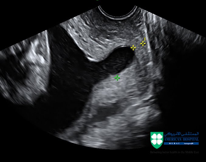
If however you opt to have your cervix(neck of the womb) measured as a screening test for preterm labour this is best done by an internal scan.
 The doctor will explain to you the type of scan that needs to be done and a chaperone will be in the room during internal scans.
Having an ultrasound scan should not be painful. If in any case you feel uncomfortable let the doctor know as he/she is usually concentrating at the ultrasound screen.
What about having a full bladder? A full bladder helps with transabdominal scans as it pushes your womb upwards and at the same time pushes the bowel out of the way creating good window to look at your baby. However you should not feel uncomfortable and be able to enjoy your scan.
Should I bring my friends and family with me? Although the scan is usually a very pleasant experience still the doctor need to concentrate as it is an important medical examination. In most cases it is best if your partner or a friend accompanies you at the scan room rather than a large number of people.   

What information will I get?

At the end of the scan YOU SHOULD BE GIVEN A COMPUTERIZED REPORT which will describe the structures seen and if there were any particular difficulties during the scan. THIS IS A VERY IMPORTANT PART OF YOUR ANTENATAL RECORD AND IT IS VERY USEFUL TO HAVE A COPY.
At this stage a large number of structural problems that a baby may have can be picked up. Ultrasound can usually provide information as to how various organs are put together and not how well they function.
Please note that scans are not foolproof. They do not show all the anomalies and in some occasions anomalies can be missed. A normal scan is not a guarantee of the delivery of a perfect baby.
Sometimes it is difficult to get good views of the baby. Common reasons for this is the baby’s position, an overweight mother or scar tissue from previous operations. In such cases the scan might need to be repeated another day.
Usually we will be able to comment on the baby’s gender at this stage. Please make sure that you let the doctor know if this is something that you want or do not want to know before the scan starts. Although doctors and sonographers are pretty good in finding the baby’s sex at this stage you should be aware that
  1. It is not 100% accurate
  2. It is not one of the primary aims of the scan (this is to confirm that major structures are normal) and it might not be possible to be seen during the allocated time. 

What are the risks for my pregnancy?

Ultrasound scanning has been routinely used in obstetrics for more than 20 years. Routine scanning does not pose any risk to your baby or yourself. 

What if there is a problem?

Most ultrasound scans are happy experiences but sometimes problems are either seen or suspected. At this stage of the pregnancy possible problems that can be picked up are
  1. Suspicion or evidence of a structural anomaly (problems relating to how the baby has developed). The doctor will explain the findings to you and depending on the problem explain what are the implications for you pregnancy and the baby. If you are having your scan by a sonographer they should be able to inform you of their concerns and arrange an urgent consultation with your obstetrician. In most cases your doctor after explaining the initial findings will arrange for you to see a Fetal Medicine Specialist. In some occasions further tests may be needed to clarify the diagnosis.
  2. A low lying placenta. Although this is not a problem at this stage of the pregnancy further scans will be needed later in pregnancy to confirm that the placenta has moved out of the way. Ask the doctor to provide you with the relevant information sheet.

  What if I still have questions about my scan?

Do not feel afraid to ask questions about aspects of the scan or the report that you do not understand. Your doctor should be able to provide you with clear information or arrange further consultation with a specialist.  

Dr George Michailidis, MRCOG
Fetal & Maternal Medicine Specialist
Consultant Obstetrician & Gynaecologist


Leave a comment

What is the role of a Fetal & Maternal Medicine Specialist – Perinatologist – in the care of a "high risk" pregnancy?

For most women, pregnancy is asmoothand incredible journey leading to the birth of their baby.However, some mayneedadditional preparation before they become pregnant, and a closer follow up during their pregnancy.These pregnancies are commonly called “high risk”. Fetal and Maternal Medicine has developed as a subspecialty of Obstetrics in order toprepareobstetricians and equip them with the appropriate skills to care for thesehigh riskpregnancies.

Fetal & Maternal Medicine Specialists, also known asperinatologists, are obstetricians who specialize in diagnosis andcareof women with complicated pregnancies. The services they provideincludepreconception counseling, detailed ultrasound examinations, fetal and maternalsurveillance. Multiple pregnancies, pregnancies complicated by diabetes or autoimmune diseases like SLE are typical examples of cases that are referredto aperinatologist. Fetal Medicine Specialists are the ones who will provide the initial counseling when fetal abnormalities are diagnosed.They have been trained to perform a number ofstateof the art diagnostic and therapeutic procedures while the baby is still in the womb.

Mothers will usually be referred to a Perinatologist by their obstetrician. The Fetal & Maternal Medicine Specialist works alongside with the mother’s obstetrician and helps to develop a personalized plan of care for each pregnancy. They also play aleadingrole in coordinating thecareof complex cases and ensuring appropriate arrangements are in place for thebaby’s birth. For example liaising with the neonatologist who will look after the baby after birth, the pediatric surgeon or pediatric cardiologist.

Being referred to a Fetal Medicine Specialist is not a reason to be alarmed or worried. In most cases is doneas a precaution in order to achieve the best plans for the pregnancy care. Some parents maychooseto see a specialist without referral from their obstetrician.

Dr George Michailidis, Obstetrician and Gynaecologist 
Fetal and Maternal Medicine Consultant 

Leave a comment

Parvo Virus B19 infection during pregnancy; What to do in Dubai.

Parvo  virus (B19) information

Schools have started again in Dubai. The large concentration of children in one place creates conditions for easy transmission of some infectious diseases. In general this is part of our natural exposure to infections that builds up our immune defence.
However there are some infections that if you have them while pregnant can affect your unborn baby. The two most widely known are rubella ( German measles) and chicken pox.  Almost all women are checked during their pregnancy regarding their immunity for these diseases so they are alerted and they have a plan of what to do if they are not immune. Another common childhood disease is the “Slapped cheek” or “fifth disease”. Having an infection with Parvo Virus B19 while pregnant can cause complications to your pregnancy. Here are some basic facts;
·Parvo B19 is a DNA virus. It is responsible for the “slapped cheek” or “fifth disease”
·Usually (in European countries) there are spring autumn epidemics that seem to come in four year cycles.
·The incubation period (the time from when you get infected to when you show the first symptoms) is usually 13-18 days while the period of maximum infectivity is during days 7-10. The risk of infection is greater to those that have frequent contact with children; teachers, day care workers, women with school age children.
·Children and adults with normal immune system usually have mild symptoms; rash, arthropathy (joint pains) Infection results in lifelong immunity.
·About 35–45% of women of childbearing age do not possess protective IgG antibodies against B19V and are not immune.
·If the mother is infected the virus can affect the baby. In particular it destroys red cell precursor cells- the cells responsible for the production of red blood cells and it can affect the fetal heart (can affect the heart muscle).
·If a mother gets infected there is a 4% risk for the baby to develop severe anaemia and hydrops. (this is when a baby presents with skin swelling and fluid collection in its chest and abdomen/tummy)
·The most difficult period is that of 8-20 weeks when the baby relies on its liver to produce red blood cells ( risk for hydrops 8% )
·
    This is preventable and treatable. Special ultrasound scans  at Fetal Medicine Unit can check whether your baby shows signs of anaemia (Doppler studies of the Middle cerebral artery). For those who have already significant symptoms a in utero fetal transfusion can be life saving.
What to do if a mother comes in contact with someone who is known to have parvo virus.
Your doctor should check your antibodies (virology tube).
·If she is IgG positive, IgM negative  reassure- evidence of immunity
·If she is IgG positive, IgM positive refer to Fetal Medicine specialist as this is evidence of recent infection.
·If she is IgG negative, IgM positive you have to be referred to a Fetal Medicine Specialist. This is evidence of recent infection but the mother may still be infective, so she will need arrangements for separate scan and to be advised to avoid meeting  with other pregnant mums.
·If she is IgG negative and IgM negative she is not immune. Advise to avoid possible contact . Repeat bloods in 7-10 days to exclude very recent infection.
Usually IgM antibodies will develop within 7 days and persist for 2-4 months.
Mothers who are referred to our Fetal Medicine Unit will be followed with serial scans looking for possible evidence of anaemia.
Please be aware that reduced fetal movements are a common manifestation of significant fetal anaemia.

Dr George Michailidis, Obstetrician and Gynecologist 
Fetal and Maternal Medicine Consultant 

1 Comment

What you need to know about premature labor and delivery in Dubai.

What you need to know about premature labor and delivery in Dubai.
What is premature (preterm) birth ?
Premature or preterm birth (delivery) is defined as a birth that takes place before 37 completed weeks of pregnancy. Unfortunately preterm labour is a relatively common pregnancy problem, up to 12.7% of births in the US were premature . Although some of preterm births are due to medical intervention the majority (75%) is due to spontaneous preterm labor. Preterm delivery is usually subdivided it in the following groups;
Late preterm birth when it occurs between 34-36 weeks
Moderately preterm birth when it occurs between 32-34 weeks
Very preterm birth when it occurs between 28-31 weeks
Extremely preterm when it occurs before 28 weeks
APPROXIMATELY 2% OF ALL PREGNANCIES DELIVER BEFORE 32 WEEKS.
Significance of preterm birth.
Up to 50% of deaths in babies born without abnormalities are related to problems caused by been born prematurely (CEMACH 2000 -2003). The following is the neonatal death rates in the UK as it was reported at the 2008 CEMACH publication.
Warning signs of Preterm labor
the following are common complains of women who eventually get in preterm labor.

□Increase in the amount of vaginal discharge. Usually the discharge changes in type and becomes watery or sometimes bloody.
□Pelvic or lower abdominal pressure.
□Constant, low, dull backache
□Mild abdominal cramps.
□Regular or frequent contractions or uterine tightening, often painless
□Ruptured membranes (your water breaks with a gush, or sometimes even a trickle of fluid)

What are the common risk factors?
this is a useful list that appears at the ACOG website Risk Factors for Preterm Labor
Check any box that applies to you:
□You have warning signs of preterm labor.
□You have had preterm labor during this pregnancy.
□You had preterm labor or preterm birth in a previous pregnancy.
□You smoke cigarettes or use cocaine.
□You are carrying more than 1 baby.
□You have an abnormal cervix (due to surgery, for instance).
□You have an abnormal uterus.
□You have had abdominal surgery during this pregnancy.
□You have had an infection while pregnant.
□You have had bleeding in the second or third trimester of your pregnancy.
□You are underweight.
□You had a mother or a grandmother who took DES (diethylstilbestrol, a medication given to many pregnant women in the 1950s and 1960s).
□You have had little or no prenatal care.
□You have a child with chromosomal disorders.
If you checked any of these boxes, you may be at risk for preterm labor. Despite what is known about these risk factors, much remains to be learned about preterm labor. Half of the women who go into preterm labor have no known risk factors.
Screening for premature labour.
A comprehensive history will should alert your obstetrician regarding the risks for preterm  birth. Unfortunately most of cases of preterm birth occur in women with no obvious risk factors.
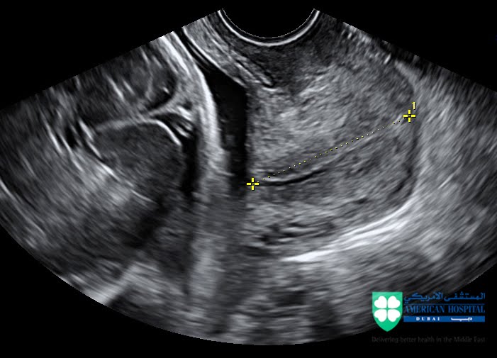
Measuring the length of your cervix is a powerful tool in indentifying those women at risk for preterm birth. This is especially important for pregnancies before 34 weeks when the impact to the baby’s wellbeing is greatest.
The mean measurement for the cervical length at 22-24 weeks is 36 mm. A short cervix <15mm is found in 1% pregnancies and about 30% of them will deliver before 34 weeks.
THE CERVIX SHOULD BE MEASURED BY A TRANSVAGINAL (INTERNAL) SCAN. Abdominal (through your tummy ) scan although can alert in case of an opening cervix does not produce good quality measurements.


The Fetal Medicine Foundation offers a comprehensive video lecture that deals with the role of screening for preterm labour by measuring the cervical length. 
MAKE SURE YOU OBTAIN A WRITTEN REPORT of the finding as well as images that demonstrate the cervical length measurement. This is essential for tracking the changes of you cervix.
Prevention and treatment options.
The purpose of screening for preterm labour is to offer interventions that can prevent or at least reduce the risk for extremely and very preterm labour and delivery. Working with a good obstetrician will ensure that you draft the best management plan for your pregnancy. This should be personalised to suite your needs and be based on the best evidence.
A few points worth mentioning ;
  • Traditionally bed rest was advocated as a way of reducing the risk for preterm birth. Unfortunately till now no major study has shown any significant advantage in strict bed rest regimes. On the contrary there are serious risks directly associated with bed rest in pregnancy like deep vein thrombosis. Discuss your life style with your doctor who should be able to give you good advice regarding your daily activities based on your case review.
  • Cervical cerclage.  Cervical cerclage has its place in selected cases. By including ultrasound screening a good obstetrician should be able to reduce the need for unnecessary interventions.
  • Progesterone supplements; This is the only intervention that has shown good results in preventing extreme preterm labour in singleton pregnancies.

Your doctor after assessing carefully the factors mentioned should be able to provide you with the best care plan for your pregnancy.

Dubai realities and premature labour
There are a number of factors that especially expatriots should keep in mind in cases that are at high risk for  preterm labour and delivery.
  1. 1.Availability of neonatal intensive care beds. At the moment even the large private units only support babies after 28 weeks of gestation. The main units that can care for babies under 28 weeks are Al Wasl Hospital and Dubai Hospital. Both units are heavily overprescribed and it can be difficult to find a bed for your baby.
  2. 2.Financial issues. Even when you deliver after 28 weeks the baby or babies (in case of multiple pregnancies) will have to spent a lot of time in an intensive care unit. The cost may easily spiral out of control. Make sure you know what you insurance is going to cover. Especially for those with higher order multiple pregnancies (triplets etc) make sure you budget for the event of extreme preterm delivery.
  3. 3.Choose your obstetrician and hospital carefully.

Dr George Michailidis, Obstetrician and Gynecologist 
Fetal and Maternal Medicine Consultant 
Useful additional reading :   


1 Comment

What is a Chorionic Villus Sampling and how is it done?

Chorionic Villus Sampling (CVS) is an invasive diagnostic procedure performed during pregnancy. It helps to obtain a tiny sample of the placenta (afterbirth) which in turn is used for the diagnosis of certain genetic conditions. We regularly perform CVS procedures at the Fetal Medicine Unit of the American Hospital Dubai. It is best if you read this information and discuss it with your Obstetrician / Gynecologist prior to having a CVS procedure. 

Why should I have a CVS?
A chorionic villus sampling (CVS) is not a routine test. Your obstetrician may offer you a CVS if there are concerns about specific genetic conditions. The following are typical examples;
·The one most widely known genetic problem is Down’s syndrome. A number of screening tests are available to help parents and obstetricians determine the risk of a pregnancy being affected before deciding for an amniocentesis. The best test to date is the combined first trimester screening. This is part of the first trimester scan.
·In some cases, the test is done to exclude known genetic problems that run in the family. Examples important in Dubai – UAE  are sickle cell anemia, thalassaemia b, cystic fibrosis.
·Routine ultrasound scans on occasions pick up certain features of the baby associated with genetic conditions.
·Some parents want to be certain that their baby is not affected by a certain disorder.

How is a Chorionic Villus Sampling (CVS) done?
The procedure is usually done between week 11-14 of your pregnancy. The doctor will fully explain the procedure to you and get to sign an informed consent.


He will check your blood group, your HIV and HepB status. If you are Rh Negative you will need an injection of Anti-D after the procedure. If you are HIV or Hep-B positive having the procedure may increase the risk of passing the infection to the baby.
Usually the doctor will perform, prior to the Chorionic Villus Sampling (CVS) an ultrasound scan to examine the baby and identify the best window for the procedure.
It takes a few minutes to prepare the procedure tray. Then he will clean your tummy with an antiseptic solution. A small amount of local anesthetic will be used to num the skin where the needle is to be inserted. A thin needle is used for the local anesthetic you might find this a bit uncomfortable. Following the local anesthetic and under direct ultrasound guidance the doctor will pass a fine needle through your tummy into your womb and into the afterbirth (placenta). The doctor’s assistant using a syringe will obtain a tiny sample. It takes a few seconds to take the sample. At the end of the procedure the doctor will remove the needle.
Most women find the whole procedure uncomfortable rather than painful. On occasions you might experience mild period like pains. This not uncommon and simple paracetamol is very helpful.

What information will I get from a CVS?
This will depend on the why the test was performed.
In most cases the aim is to examine the baby’s karyotype (genetic information).
The sample taken contains cell from the placenta. Although the placenta is a separate organ from the baby it has the same genetic information. The cells obtained can be cultured in the lab. After they are cultured to increase their number and get most of them to start multiplying, using special techniques, the geneticist will look at these cells under the microscope. The karyotype is a snapshot of the baby’s chromosomes. The geneticist will count their number (they should be 46) and examine their morphology (how they look). Usually, due to the time it takes to culture the cells, this test is ready in 14 days from when it reaches the lab.
Some more specialized genetic tests need to be performed it might take longer. The doctor should be able to let you know of the approximate time before the test is done.
Rapid tests that only look at certain chromosomes in a small number of cell s (FISH- PCR) are available. These tests usually take 48h after the fluid reaches the lab. Our advice to all is to have the full karyotype in addition to the rapid tests. 
These tests in the vast majority of cases will give a definitive answer whether the baby is affected or not from the disorder you were testing for.
Please note the following;
  • The test will give a result for a specific condition. It does NOT guarantee that the baby does not have other genetic disorders.
  • Rarely while the test was performed to exclude one problem it picks up another unexpected one. Your doctor will help you to understand the results and their implications for your baby.
  • Rarely (less that 1/100) the test may not give a clear a result. A repeat test may be necessary.
What are the risks for my pregnancy
Unfortunately a Chorionic Villus Sampling (CVS) is not without risks. It is generally quoted that there is a risk of 1-2% to have a miscarriage as a result of the test.
In a small number of cases (1:1000) a serious infection might develop.
You should carefully consider these risks against the value of the information you seek to get from the test

I hope you find this information useful. For further queries do not hesitate to contact us


Dr George Michailidis, Obstetrician and Gynecologist 
Fetal and Maternal Medicine Consultant 

Leave a comment

Amniocentesis services in Dubai

Amniocentesis is a diagnostic procedure commonly performed at the Fetal Medicine Unit of the American Hospital Dubai. In this post you will find useful information that you should read and is best to discuss with your obstetrician during the preparation for an amniocentesis.

What is an amniocentesis?

Amniocentesis is a diagnostic procedure performed during pregnancy. It helps to obtain a small sample of amniotic fluid (the fluid around the baby) which in turn is usedfor the diagnosisof certain genetic conditions or infections.

Why should I have an amniocentesis?

An amniocentesis is not a routine test. Your doctor may offer youan amniocentesis if there are concerns about specific genetic conditions. The following are typical examples;
  • The one most widely knowngenetic problem is Down’s syndrome. A number of screening tests are available to help parents and obstetricians determine the riskof a pregnancy being affectedbefore deciding for an amniocentesis. The best test to date is the combined first trimester screening. This is part of the first trimester scan.
  • In some cases, the test is doneto excludeknown genetic problems that run in the family.
  • Routine ultrasound scans on occasions pick up certain features of the baby associated with genetic conditions.
  • Some parents wantto be certainthat their babyis not affected by a certain disorder.
  • An amniocentesis may also be offered to check whether an infectionhas passed from mother to the baby.


How is anamniocentesis done?

The procedure is best done after week 15 of your pregnancy. The doctor will fully explain the procedure and get you
to sign an informed consent.

 
He will checkyour blood group, your HIV and HepB status. If youare Rh Negativeyou will need an injectionof Anti-D after the procedure.If youare HIV or Hep-B positive, having the procedure may increase the risk of passing the infectionto the baby.


Usually the doctor will performprior to the amniocentesis an ultrasound scanto examinethe babyand identifythe best window for the procedure.
It takes a few minutes to prepare the proceduretray. Then he will clean your tummywith an antiseptic solution. A small amount of local anaesthetic will be usedto numb the skin where the needle is to be inserted. A thin needle is usedfor the local anaesthetic youmight find this a bit uncomfortable.

Following the local anaesthetic and under direct ultrasound guidance, the doctor will passa fineneedle through your tummyinto your womb and the fluid around the baby. The doctor’s assistantusing a syringe will obtainabout 15mls of this fluid. The babywill not miss this fluidand will replace it in a few hours. As a verythin needle is used, it takes a few seconds to takethe sample. At the end of the procedure, the doctorwillremove the needle.

Most women find the whole procedure uncomfortablerather than painful. On occasions, youmight experience mild period like pains. This not uncommon and simpleparacetamol is very helpful.

In some occasions, not enough fluid is obtainedwith the first attempt. This is usuallydue to the position of the baby. In this case, the doctor may have to reinsert the sampling needle.

What information will Igetfrom an amniocentesis?

This will depend on the why the test was performed.

In most cases, the aim is to examine the baby’s karyotype (genetic information).
Within the fluid withdrawn, there are cells that come off the baby’s skin. These cells (unlike our outer skin cells) are alive and can be cultured in the lab. After they are culturedto increase their number and getmost of them to startmultiplying, using specialtechniques, the geneticist will look at these cells under the microscope. The karyotype is a snapshot of the baby’s chromosomes. The geneticist will counttheir number(they should be 46) and examinetheir morphology (how they look). Usually, due tothe time it takes to culture the cells, this test is readyin 14 days from when it reaches the lab.

If more specialized genetic tests need to be performed it might take longer.Your obstetrician should be able to letyouknow of the approximate time before the testis done.

Rapid tests that only look at certainchromosomes in a small number of cells (FISH- PCR) are available
. These tests usually take48h after the fluid reaches the lab. Ouradvice to all is to have the fullkaryotype in addition to the rapid tests.

These tests in the vast majority of cases will givea definitive answer whether the baby is affectedor not from the disorder youwere testing for.


Pleasenotethe following;

The test will givea resultfor a specific condition.
It does NOT guarantee that the babydoes not have other genetic disorders.

Rarely whilethe test was performedto excludeone problem it picks up another unexpected one. Your Obstetrician, sometimes with the help of a geneticist, will help youto understand the results and their implications for your baby.
Rarely (less that 1/100) the cells do not grow well in the lab and the testcannot givea result.A repeat test may be necessary.

What are the risks for my pregnancy?

Unfortunately, the amniocentesis is not without risks. It is quotedthat there is a risk of 1% having a miscarriage as a result of the test. In ourexperience, this riskhas been much smaller (about 0.5%)

In a small number of cases (1:1000), a serious infectionmight develop.

Youshould carefully consider these risks against the value of the information youseektogetfrom the test.

The team will also explain to you; How you will receive your results.
What if my results areabnormal?

Although in many of the cases, the results will come back as normalthere will be those that are going to be affected by the conditionwe were testing for.


Youshould be aware that a termination of pregnancy is only allowed in exceptional cases at the UAE. Getting authorizationcan be a lengthy process.

We hope youwill find this information useful. If youhave any questions, you can contact either Dr Michailidis or our specialist midwife.

Dr George Michailidis, Obstetrician and Gynecologist 
Fetal and Maternal Medicine Consultant 

1 Comment

High risk pregnancy

A commonly used definition for high risk pregnancy is the following; A pregnancy is characterised as high risk when the likelihood for significant complications is higher than average. How the care of these pregnancies is managed varies around the world. For the purpose of being referred to a Fetal & Maternal specialist in Dubai the following list outlines some of the common scenarios were our input can help the management of your pregnancy (Doctor’s referral form and self referral form);

Risk for
Referral for
Multiple pregnancies (twins dichorionic)
Chromosomal/structural anomalies
Preterm delivery
Growth retardation
First trimester screening for Down’s syndrome
Morphology scans
Growth scans and Doppler studies
Multiple pregnancies
(monochorionic twins)
As above plus the risk for TTTS (1/5)
It is good practice these pregnancies to be managed by a Fetal Medicine specialist.

Suspected Fetal abnormalities
Depends on the anomaly
Morphology scan, counselling 
Fetal surveillance


History of previous pregnancy by a affected congenital anomaly

Re occurrence
Preconception counselling,
First trimester and morphology scans.
Suspected fetal growth retardation

Fetal loss / or unnecessary pregnancy intervention
Serial growth scans
Fetal surveillance
Advanced maternal age

Chromosomal anomalies (like Down’s)
First trimester screening
Particular maternal medical conditions

Pre existing diabetes or GDM on insulin
Risk for congenital anomalies/macrosomia
Consider Full management by the FMU due to the frequency of scans visits it would be easier for these patients to be followed up by the FMU with aid of an endocrinologist

Hypothyroidism (treated Graves)

Risk of fetal hyperthyroidism
Fetal surveillance
Autoimmune disease (lupus , antiphospholipid syndrome, RA etg)

Risk of deterioration of the maternal condition/ growth retardation
Preconception counselling,
Fetal surveillance
Maternal cardiac disease
Risk of deterioration of the maternal condition/ fetal abnormalities
Preconception counselling,
Fetal surveillance,
Maternal surveillance with the aid of a cardiologist

Rh negative with positive antibodies

Risk for fetal anemia  -hydrops
Preconception counselling,
Fetal surveillance


Dr George Michailidis, Obstetrician and Gynecologist 
Fetal and Maternal Medicine Consultant 


1 Comment

Twin pregnancy in Dubai

Twin pregnancies (as well as higher order multiple pregnancies) have been on the rise. Medical interventions , like ovulation drugs and IVF treatment have been the main reasons for the increase in multiple pregnancies the recent decades.  The needs and the care of mothers who are carrying twins are different compared to those of a singleton (one baby ) pregnancy. Furthermore all twin pregnancies are not the same. It is very important to know the type of twins you are carrying as this has major implications in the management and care of the pregnancy.
The best time to determine the type of twins is in early pregnancy (usually 10-13 weeks). What you NEED to know is if your twins are monochorionic or dichorionic. Not so much if they are boys or girl, identical or not. Determining chorionicity is essential for the proper management of your pregnancy. Ask your doctor to make a clear documentation of your babies chorionicity. 

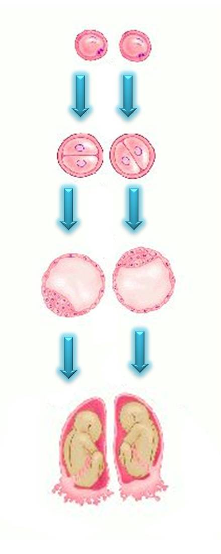
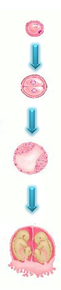
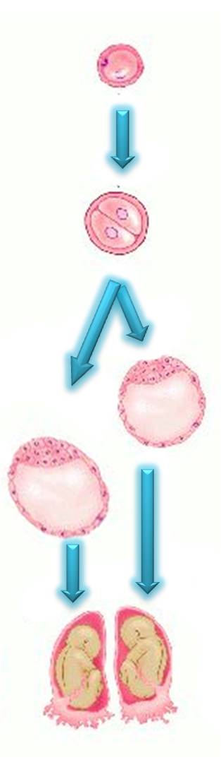
The following diagrams describe the different  type of twins.
Dizygotic (fraternal , not identical ) twins


Dizygotic (fraternal or not identical) twins are the most common type of twins (79%). Two oocytes (eggs) are fertilized by two sperm cells and form two embryos that are like brothers or sisters. These babies although similar (as siblings) are not genetically identical. They can be either the same or different gender. Each baby has each own placenta. Sometimes the two placentas fuse as they develop one next to the other. The two placentas do not communicate between  them.
These are the type of twins you most commonly have after IVF as two fertilised embryos are returned into the womb.  
All dizygotic twins are dichorionic (with few rare exceptions). 


Monozygotic (identical ) monochrionic twins 
Monozygotic dichorionic twins
Monozygotic (identical) twins. Their  frequency is about 4.5 per 1000 pregnancies. In this case one sperm cell fertilizes one oocyte (egg). The embryo that forms subsequently splits in two. These babies are genetically identical and are always the same gender. 
The most common type of identical twins are monochorionic diamniotic twins (70-80%). Imagine one balloon (the chorion) containing two smaller balloons (the amnion) each one with a baby inside. The two placentae communicate between them (blood can go from one baby to the other)

In about 20-25% when the embryo splits early (days 0-3) you have monozygotic dichorionic twins. These twins although they are genetically identical each one has each own placenta and there are no   
                                                                                                                                                       significant communications between 
                                                                                                                                                       them. 
Monozygotic monochorionic monoamniotic twins


A rare type of monozygotic ( identical) twins are those who are in the same sac. They are called monoamiotic. This happens when the embryo splits in two after day 8. Only about 2% of monozygotic twins are monoamniotic. These babies share the same placenta

If the split takes place after day 12 the results are the various type of conjoined twins.   


The reason why it is so important to determine the type of twins you carry is a syndrome called twin to twin transfusion syndrome (TTTS).  Twin to Twin Transfusion syndrome is unique to monochorionic (MC)  multiple pregnancies. It complicates up to 1/5 of MC twin pregnancies. If severe and left untreated it leads to the loss of both babies.  

Your obstetrician in Dubai should let you know from the outset the type of twins you carry and based on that draft a plan for the management of your pregnancy. 

The following are, in my opinion, important parts of the care and in our Fetal Medicine Unit we ensure we spent ample time to discuss them with the parents;
  • Screening for chormosomal anomalies
  • Nutrition and diet.
  • Monitoring for twin to twin transfusion syndrome in monochorionic twin pregnancies.
  • Risks and monitoring for preterm delivery. In Dubai it can be proven very difficult (and costly) to manage very premature babies. This is an important part of you care and should be fully discussed to ensure you reach informed choices about your care.
  • Monitoring for growth  It is good practice each time you have an ultrasound scan to get a typed report that clearly documents the findings and type of twins.
  • Timing and mode of delivery


I hope you find this helpful


Dr George Michailidis MRCOG
Specialist in Fetal & Maternal Medicine
Consultant Obstetrician & Gynaecologist


1 Comment

The value of birth plan, advice how and why you should prepare one.


Birth Plan

Birth plans used to be a terrible nightmare for young trainees in O&G in the UK including myself. We used to associate them with difficult patients, difficult decision making and sometimes conflict when the clinical circumstances forced us to deviate from the printed document. However as with many other things in life as you gain experience you recognise their real value.

As a private practitioner now I encourage my patients to consider preparing a birth plan. Our main focus is not so much the printed document but more so the “journey”  required for its preparation.
Hodnett E.  in an extensive review (Pain and women’s satisfaction with the experience of childbirth: a systematic review) concluded that the main factors influencing maternal satisfaction of their childbirth experience are the following;

1.Personal expectations.
2.Amount of support by the caregivers .
3.Quality of the patient – caregiver relationship.
4.Involvement in decision making.

A birth plan gives the opportunity to explore all of the above.
·It provides a basis for discussion that helps the medical team identify what are the key issues for the mother (personal expectations). From the mother’s perspective it  ensures that these are addressed appropriately.

·It actively involves the mother in decision making

·It provides the means of building and improving the relation between the doctor- midwife and the mother. It highlights the areas of agreement but more importantly indentifies the areas of disagreement. By doing so it  allows time to resolve misconceptions and find a “common ground” reducing the risk of conflict at the time of labour.

The following are some general tips to help you with the preparation of your birth plan.

  • Keep it simple and short. Someone will have to read it most likely in the middle of the night. The more complicated the birth plan the less likely to be followed.
  • Be pragmatic on your expectations and requests. For example dimming the lights at the time an operation may not be a good idea. What you are asking needs to be in line with the what the place you have chosen is able to offer, the hospital policies and procedures.
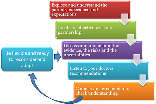
  • Be flexible and ready to adapt. It is extremely important to recognise that labour is a dynamic process. It is difficult to predict all the possible scenarios. So be ready to change and adapt if the circumstances require it.  The women who are more likely to be happy with the whole experience are those that approached childbirth with an adaptable plan.
  • After you have prepared your birth plan review it with your doctor. If you involve other people in the birth plan make sure they are aware and agree with your requests.
  • Consider the “decision making process” described  in the VBAC post  
The Canadian Society of Obstetricians & Gynecologists gives a typical example of a birth plan form.
Childbirth education is an essential element that mothers need before they embark in preparing their birth plans. There are great books, internet sites that can help women with initial information.
Midwife  led classes are an extremely valuable asset in education and preparation of parents for labour. Your doctor should be the one to help you bring everything together and approach labour with confidence.
Childbirth education should deal with the different aspects of labour. The following list is by no means exclusive but contains some of the basic things a mother should understand.
1.The process of normal labour
2.What are the common medical interventions during labour
3.How to deal with labour pain
4.When to call the hospital
5.What are the signs or symptoms that should worry you and get you talk to your doctor.
6.What to expect at the place you are planning to deliver
7.Care of the baby including breast feeding
8.Care of the mother after labour
Well respected midwife educators in Dubai are;
Ms Cecile De Scaly:
Mobile 050 6947938
Ms Hanadi Safi at Emirates Home Nursing is:
04 39633137
050 9647334
or info@emirateshomenursing.ae

Dr George Michailidis, Obstetrician and Gynecologist 
Fetal and Maternal Medicine Consultant